
Service Details
Molecular imaging by MS imaging
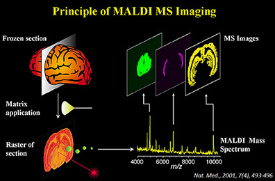
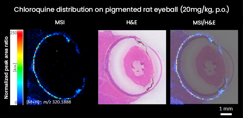
Mass spectrometry imaging (MS imaging) is a molecular imaging technique utilizing mass spectrometry. It enables imaging and evaluation of objective low molecular compounds distributed in small local areas.
Two-dimensional distribution maps are created from signal intensity and location information of the specific ions. It is possible to analyze more than two compounds such as metabolites and marker molecules at the same position.
Because this method can directly determine the compound concentrations in tissues without labelling agents such as radioisotopes or fluorescent dyes, it is expected to be utilized in drug development to visualize pharmacokinetics in target tissues.
We provide contract analytical services using FT-ICR*1 type mass spectrometry MALDI*2 imaging system (solariX 2xR) from Bruker.
*1:FT-ICR: Fourier transform ion cyclotron resonance
*2:MALDI: Matrix-assisted laser desorption ionization
Site-specific analysis by microdissection
Microdissection is a technique utilizing a dedicated laser irradiation device to recover cell groups in specific regions in tissue sections on a slide by cutting with a laser beam. For example, by cutting a selected tumor part from a histopathology section, comparing and analyzing normal and tumor parts can be conducted.
Recovered tissue samples are used to determinate concentrations of target compounds and gene expression analyses by LC-MS, PCR, and sequencer. Compared with ordinary analyses with heterogeneous cell groups, analyses with homogeneous cell groups are expected to reflect the character of the cell groups more.
Spatial profiling by GeoMx
Mediford Corporation is working on spatial profiling analysis that combines imaging technology and quantitative analysis of gene expression and proteins.
With GeoMx Digital Spatial Profiler from Bruker Spatial Biology we can specify a Region of Interest (ROI) in the fluorescence microscope image of a tissue section slide and quantitate the gene expression and proteins in that region. Probe or antibodies for detecting target RNA and proteins are labeled with oligonucleotide barcodes, and the barcodes in the specific region are released by UV irradiation and detected/quantitated with nCounter Analysis System from Bruker Spatial Biology and NGS system from Illmumina.


Histological stain/preparation of virtual slides
Immunohistochemistry (IHC) is a technique which visualizes target antigens by antigen-antibody reactions on tissue samples retaining their microstructures.
It is used for pathological diagnosis in non-clinical and clinical areas, and also used to evaluate expressions of specific markers in drug development.
Histopathologic inspection in clinical areas is contracted by LSI Medience. Mediford Corporation conducts histopathologic inspection in non-clinical areas and expression analysis of unique markers which is out of the scope of clinical diagnostic testing.
We provide series of services such as preparation of tissue blocks/slides, histological stains, preparation of virtual slides, etc.
